Specifications
Histology of Domestic Animals Slide Set II includes 24 microscope slides:
- Apis mellifica, honey bee, mouth parts of worker w.m.
- Apis mellifica, honey bee, posterior leg with pollen basket w.m.
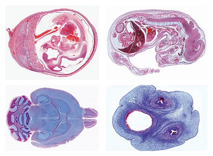
- Brain of mouse, horizontal l.s. of the complete organ
- Cerebellum, t.s. stained by Golgi’s silver method to show the Purkinje cells
- Fallopian tube of pig, t.s.
- Hair (bristle) of pig, w.m.
- Kidney of rabbit, t.s.
- Liver of pig, t.s.
- Mammary gland of cow, active t.s.
- Merkel corpuscles in t.s. through snout of pig
- Olfactory region from nose of rabbit, t.s.
- Ovary, in t.s. of abdomen of queen, Apis mellifica
- Ovary of cat, t.s. for general study, shows primary, secondary and Graafian follicles
- Peripheral nerve of cow or pig, l.s. routine stained
- Retina of pig, thin sec. special stain for details of rods and cones
- Skin of foot, cat, vertical sec. showing stratum corneum and stratum germinativum
- Sperm smear of bull
- Spinal cord of cow, t.s. stained for Nissl bodies
- Taste buds, t.s. of papilla foliata in tongue of rabbit shows abundant taste buds, carefully stained
- Testis and epididymis of cat, t.s.
- Testis, in t.s. of abdomen of drone, Apis mellifica
- Uterus of pig, pregnant stage, t.s.
- Uterus of pig, resting stage, t.s.
- Young mouse, sagittal l.s. through entire specimen passing the vertebral column